Imbricatea Cavalier-Smith 2012
Cells with secreted surface silicious scales.
Cells with secreted surface silicious scales.
1.0 Euglyphida Copeland 1956, emend. Cavalier-Smith 1997
Test of organic material; most taxa with secreted silica scales held together by an organic cement.
1.1. Trinematidae Hoogenraad & De Groot 1940, emend Adl et al. 2012
Test with bilateral symmetry; scales oval or round, sometimes of both types; specialized tooth-shaped scales around the aperture; aperture invaginated in some taxa.
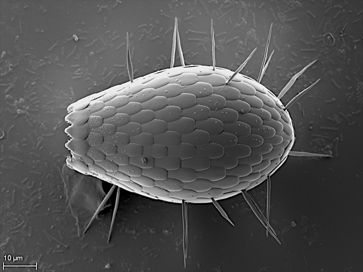
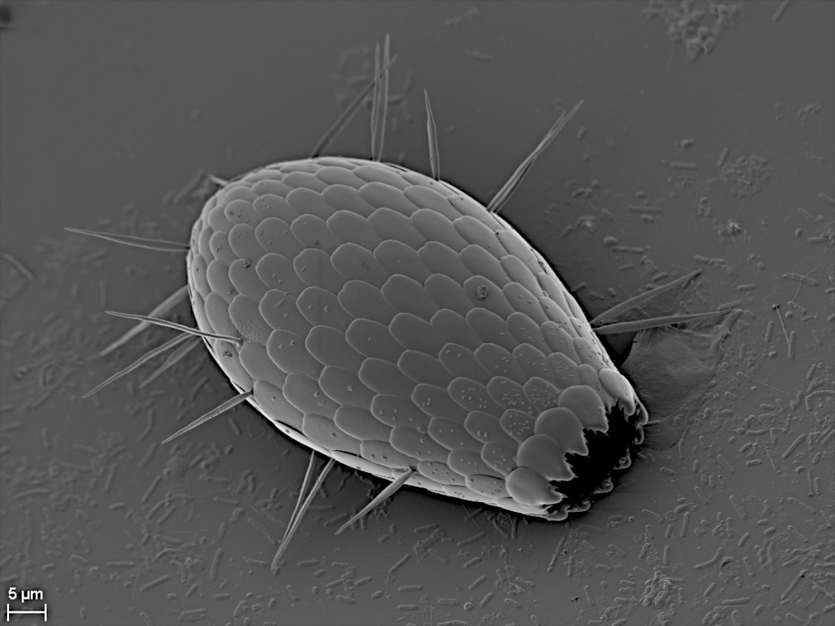
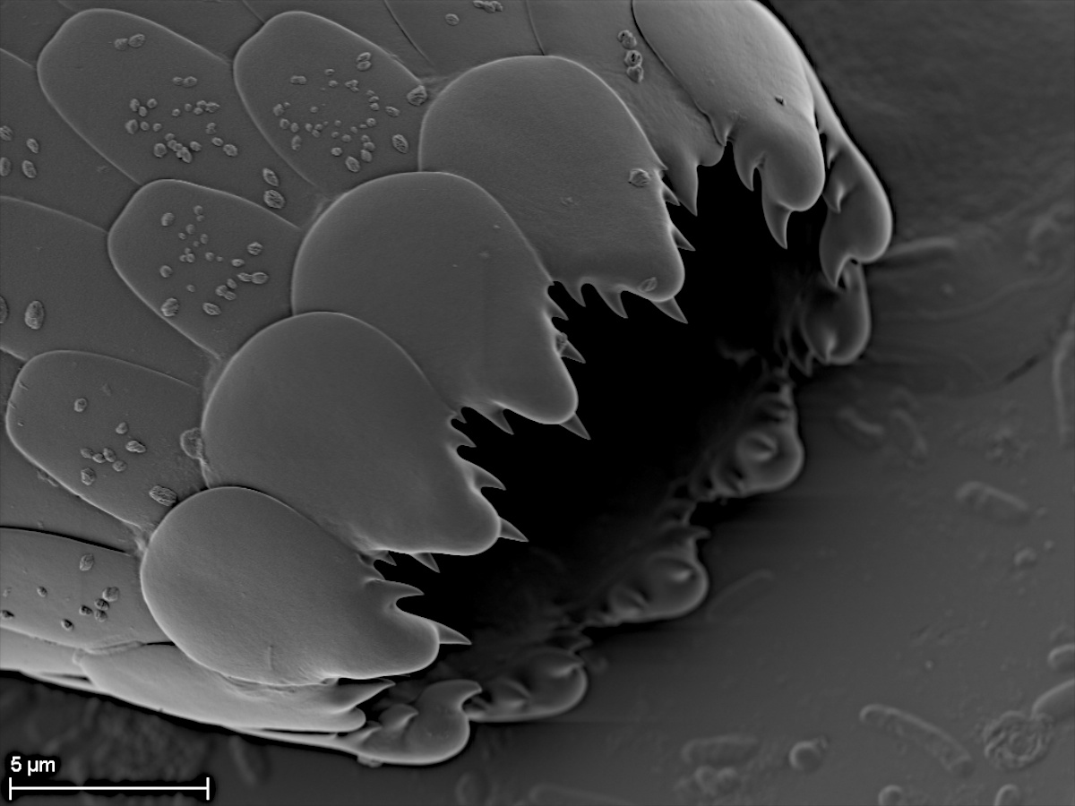
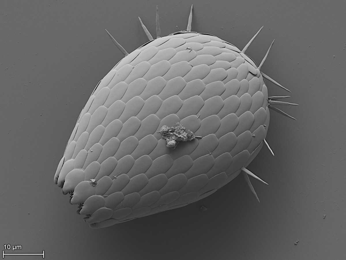
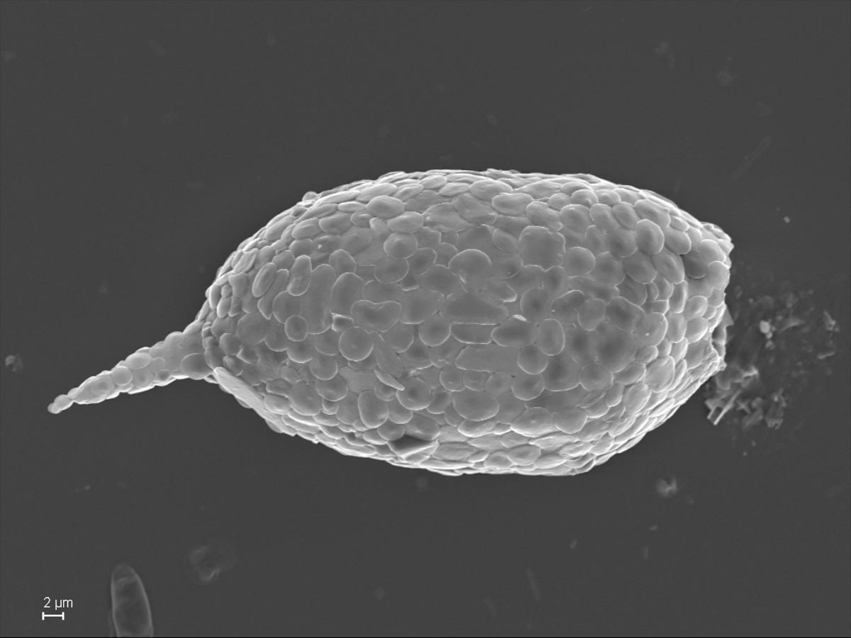
1.1.1. Trinema Dujardin 1841
Test with large circular scales and smaller circular or ellipsoid scales; with tooth-shaped apertural scales.
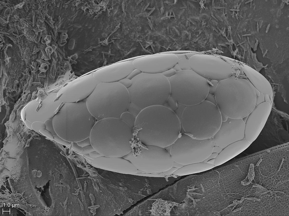
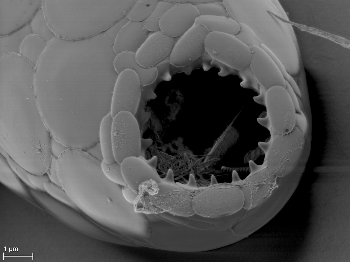
Trinema sp.
Trinema sp.
1.2. Euglyphidae Wallich 1864, emend Lara et al. 2007
Thin, overlapping, elliptical scales; presence of specialized scales around the aperture with typical indentation.
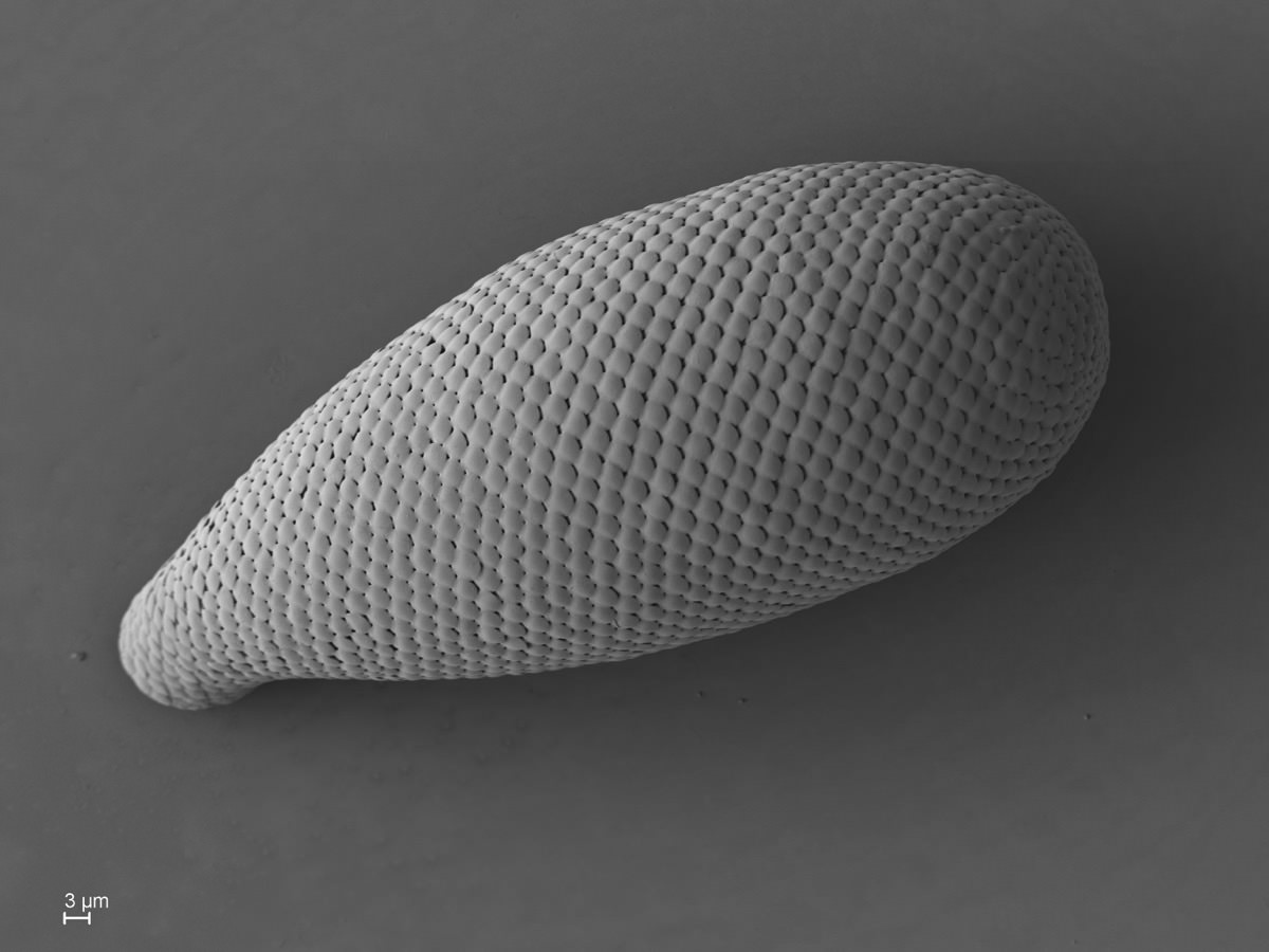
1.2.1. Euglypha Dujardin 1841
Test elongate ovoid or pyriform; aperture with denticulate mouth pieces.
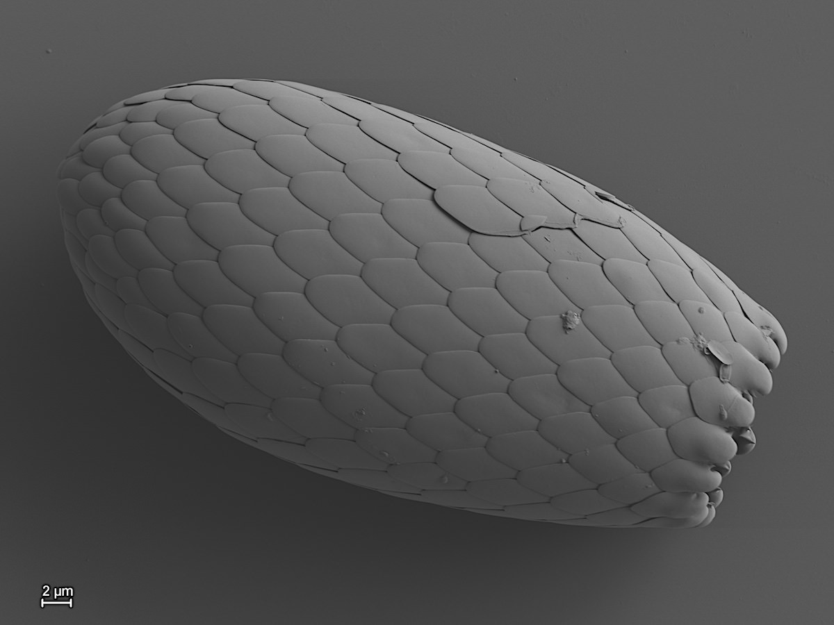
Euglypha rotunda Wailes 1911
Euglypha compressa Carter 1864
Euglypha sp.
1.2.2. Pareuglypha Penard 1902 (incertae sedis)
Test ovoid, tapered towards a circular aperture. With a scaly spine of variable length.
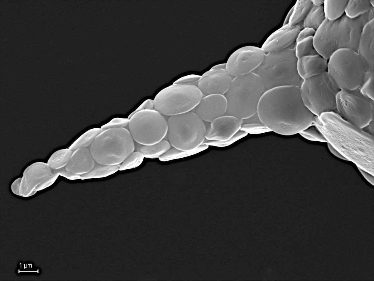
Pareuglypha reticulata Penard 1902
1.3. Cyphoderiidae de Saedeleer 1934
Scales circular, oval or kidney-shaped, juxtaposed or imbricated; test aperture angled, some with collar.
1.3.1. Cyphoderia Schlumberger 1845
Test retort-shaped; colorless to yellow; made up of a thin chitinous membrane, covered with discs or scales; aperture terminal, oblique, circular; body does not fill the test completely; nucleus large, posterior; pseudopodia, few, long filose, simple or branched; fresh water.
Cyphoderia sp.
1.4. Sphenoderiidae Chatelain 2013
Self-secreted scales, circular or elliptical; aperture surrounded by small round or oval scales.
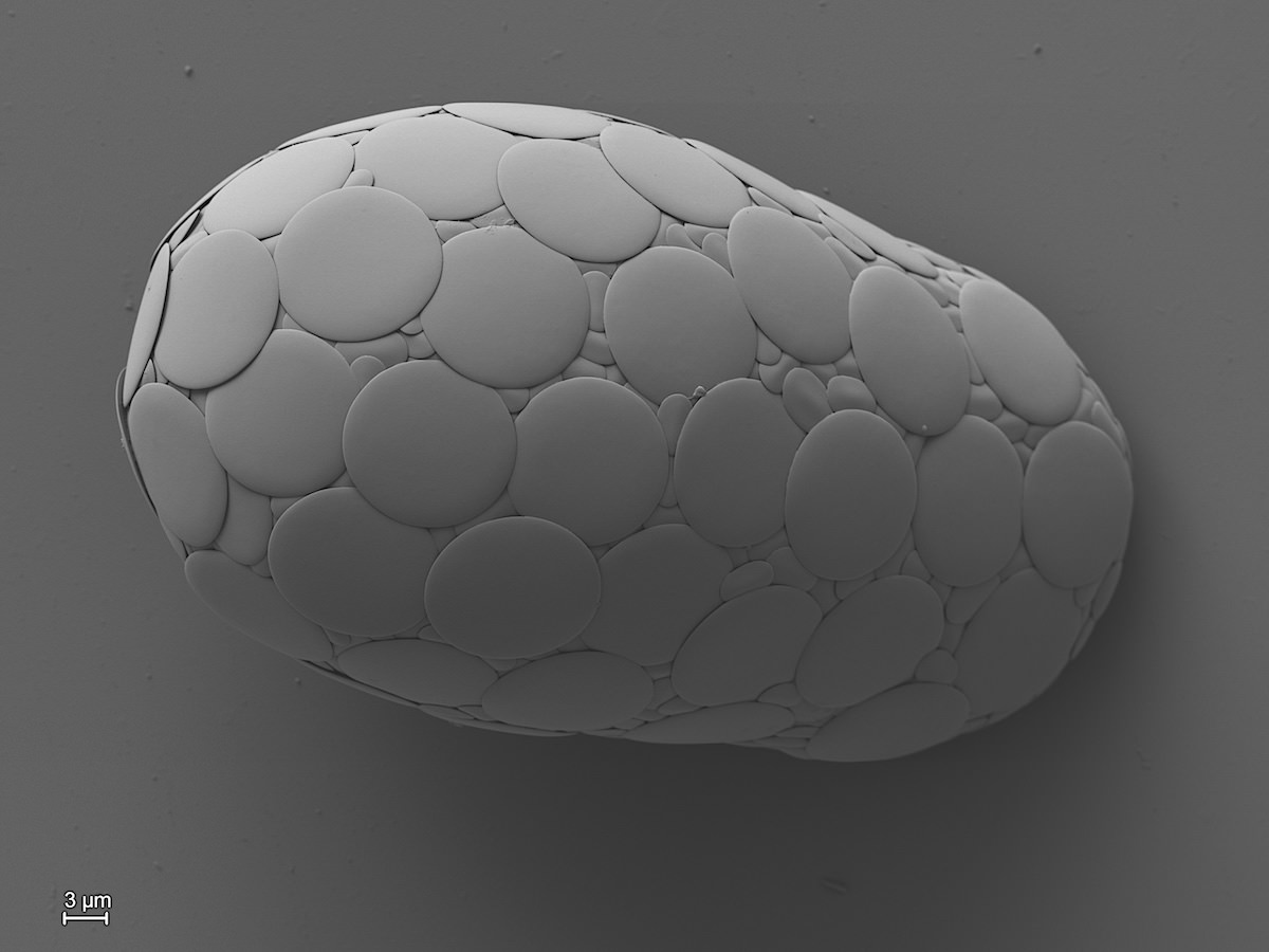
1.4.1. Sphenoderia Schlumberger 1845
Test clear, circular or ovoid, covered with overlapping elliptical or nearly circular scales. Aperture circular or a narrow, oval slit at the end of a broad, clear collar covered with numerous small plates. Nucleus spherical, with few small nucleoli.
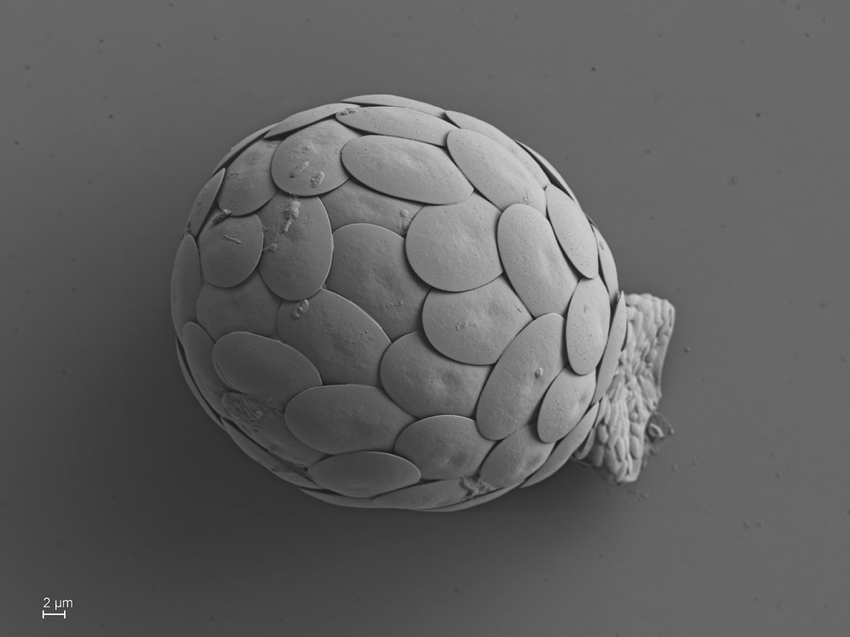
Sphenoderia lenta Schlumberger 1845
2.0. Placoperla Cavalier-Smith 2012
Imbricate cercozoa that secrete a test of mineralized spherical pearls or silicified plate scales that are typically double tiered with a perforate upper tier.
2.1. Thaumatomonadida Shirkina1987
Heterotrophic usually gliding cells that may swim also; with flattened cell body and with two heterodynamic cilia inserting subapically and/or ventrally; some unikont; with extrusomes; filopodia produced subapically or from ventral groove; cysts; multinucleate and multiciliate stages known.
Heterotrophic usually gliding cells that may swim also; with flattened cell body and with two heterodynamic cilia inserting subapically and/or ventrally; some unikont; with extrusomes; filopodia produced subapically or from ventral groove; cysts; multinucleate and multiciliate stages known.
2.1.1. Thaumatomonas de Saedeleer 1931
Bicilated, heterotrophic amoeboflagellates with silicious body scales.
Bicilated, heterotrophic amoeboflagellates with silicious body scales.
Thaumatomonas sp.
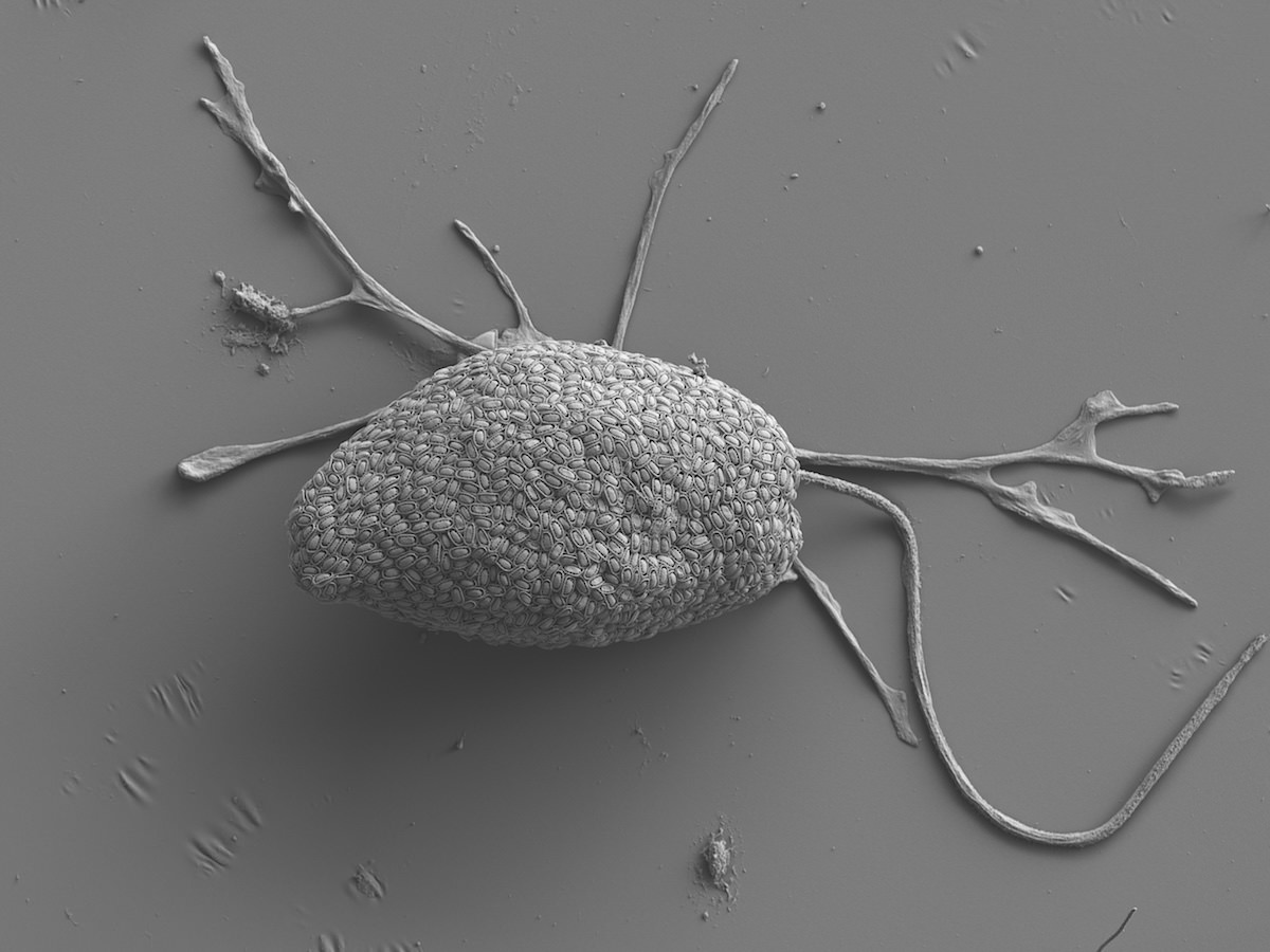
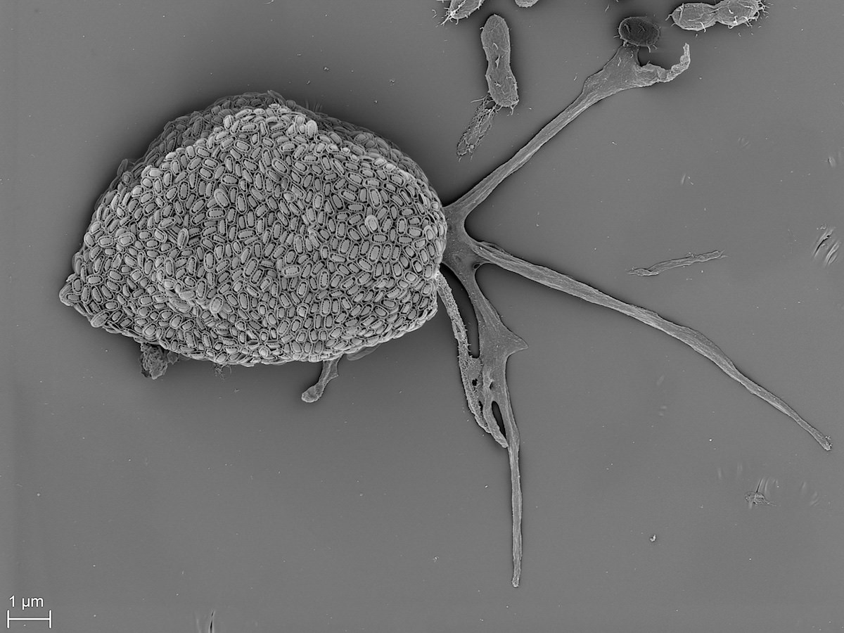
Thaumatomonas sp.
2.2. Perlatia Cavalier-Smith 2012
Imbricate cercozoa that secrete a test of mineralized spherical pearls.
3.1.1. Perlofilida Cavalier-Smith 2012
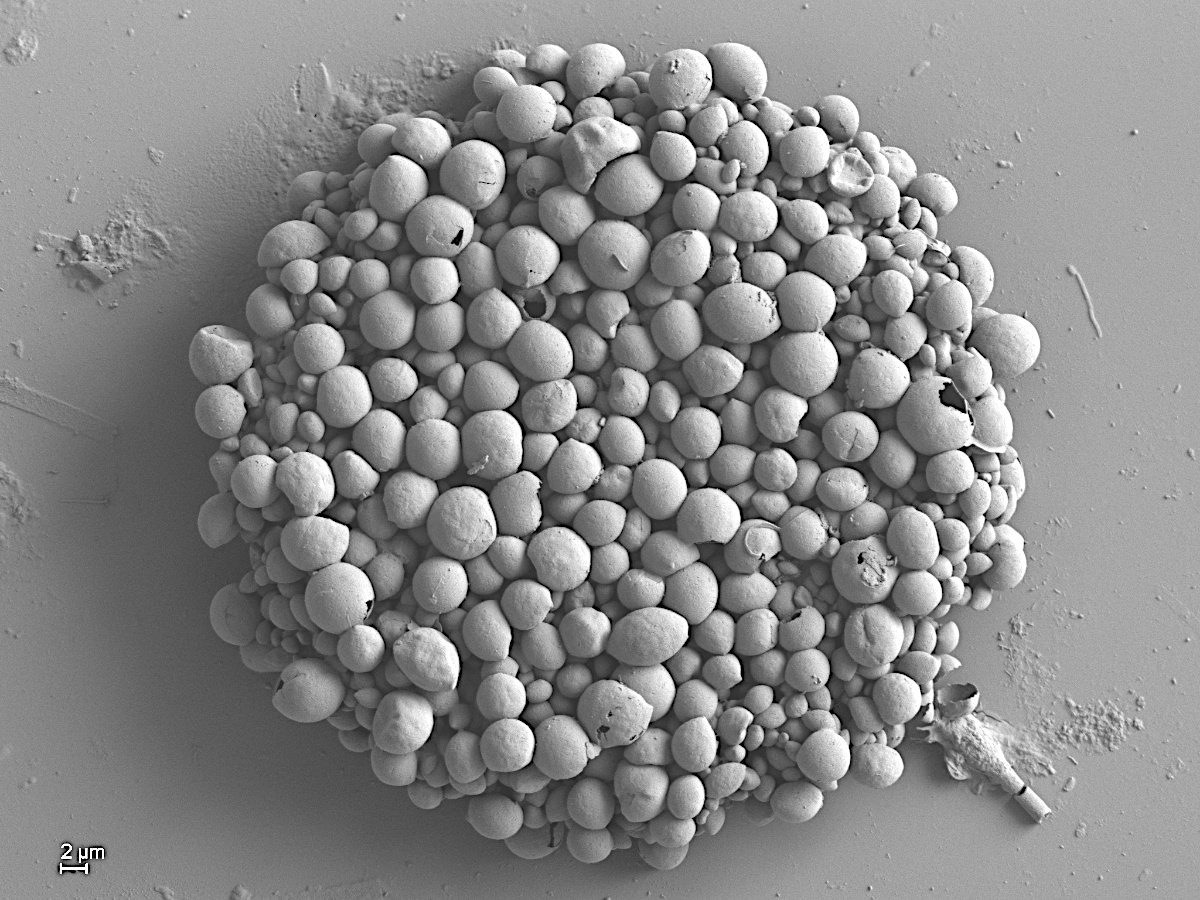
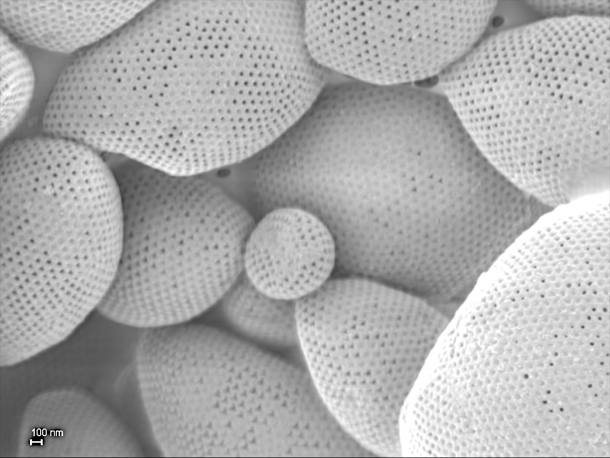
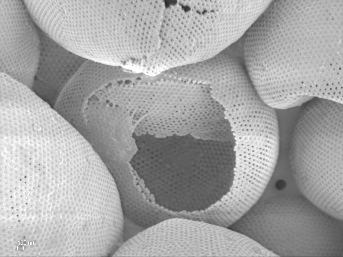
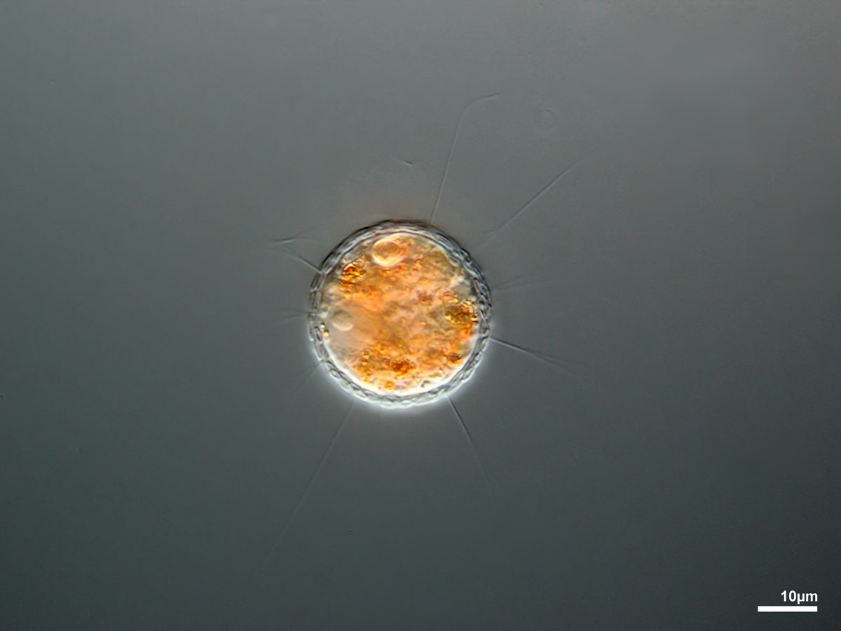
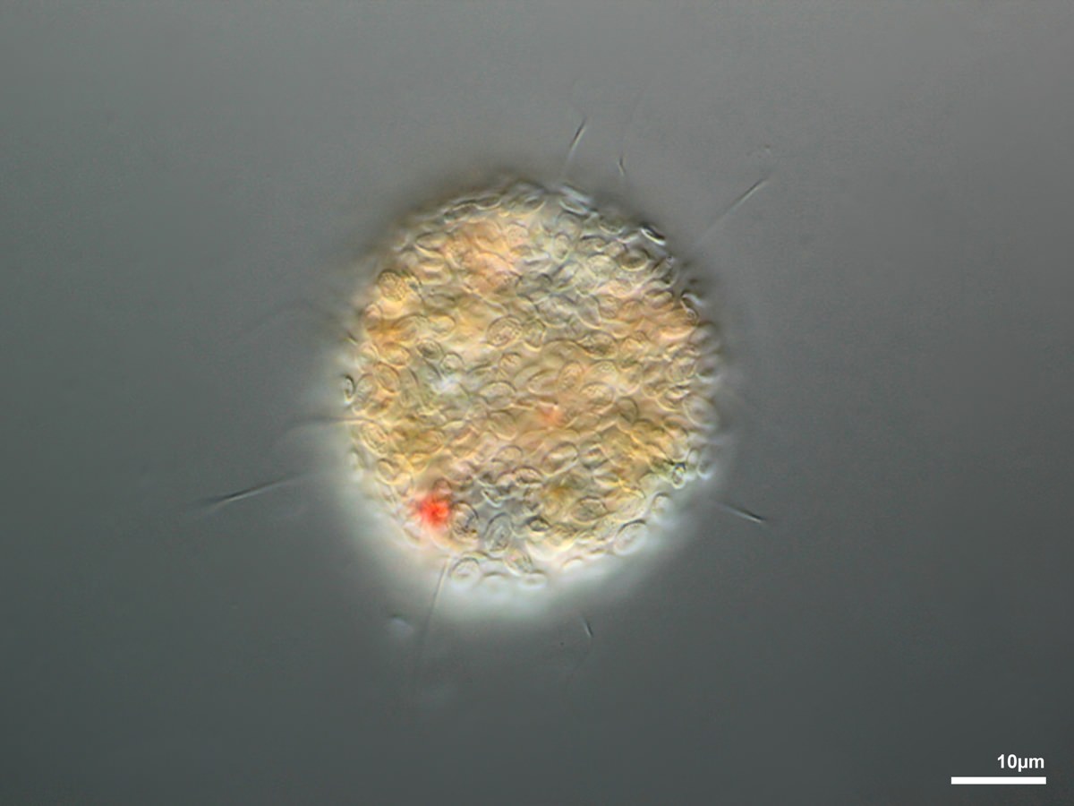
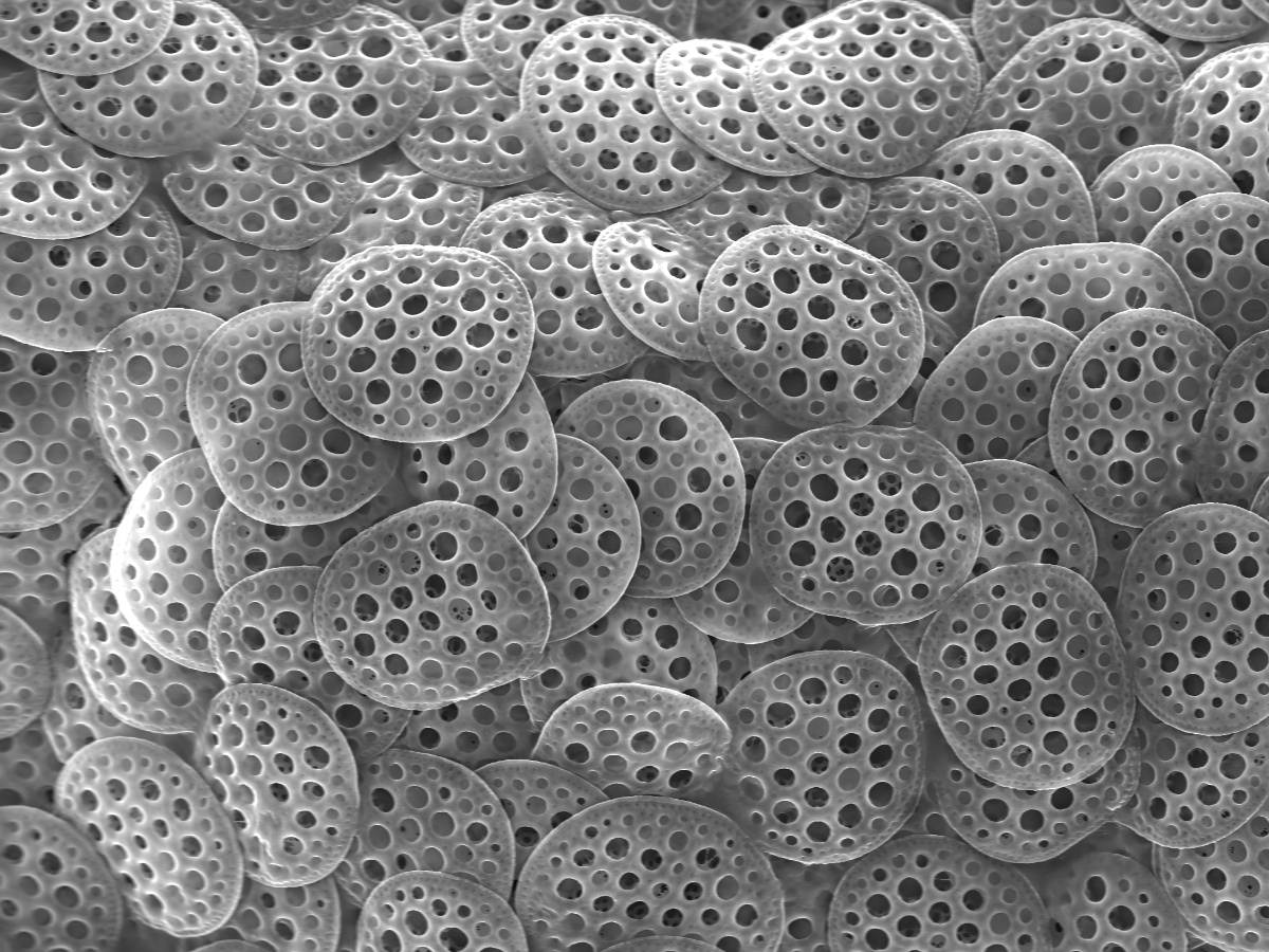
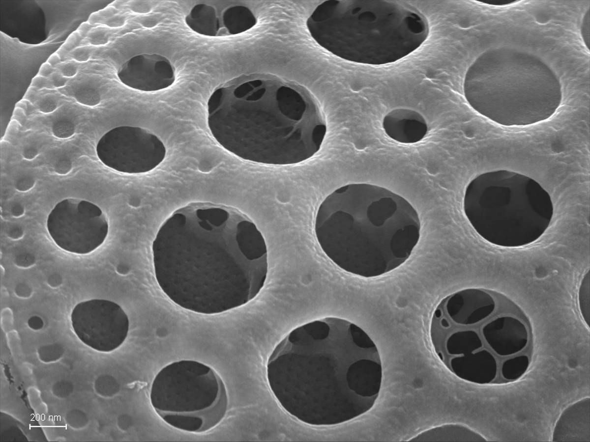
Near-spherical heterotrophic and phagotrophic non-flagellate cells with uniformly extremely thin but stiff, symmetrically radiating, unbranched filopodia, cell surface covered with one or more layers of spherical siliceous pearls, bounded by a wall with numerous uniformly sized but irregular-shaped small holes.
3.1.1.1. Pompholyxophryidae Page 1987 (incertae sedis)
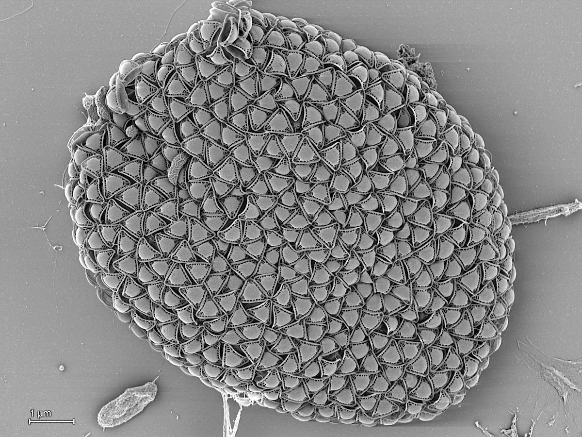
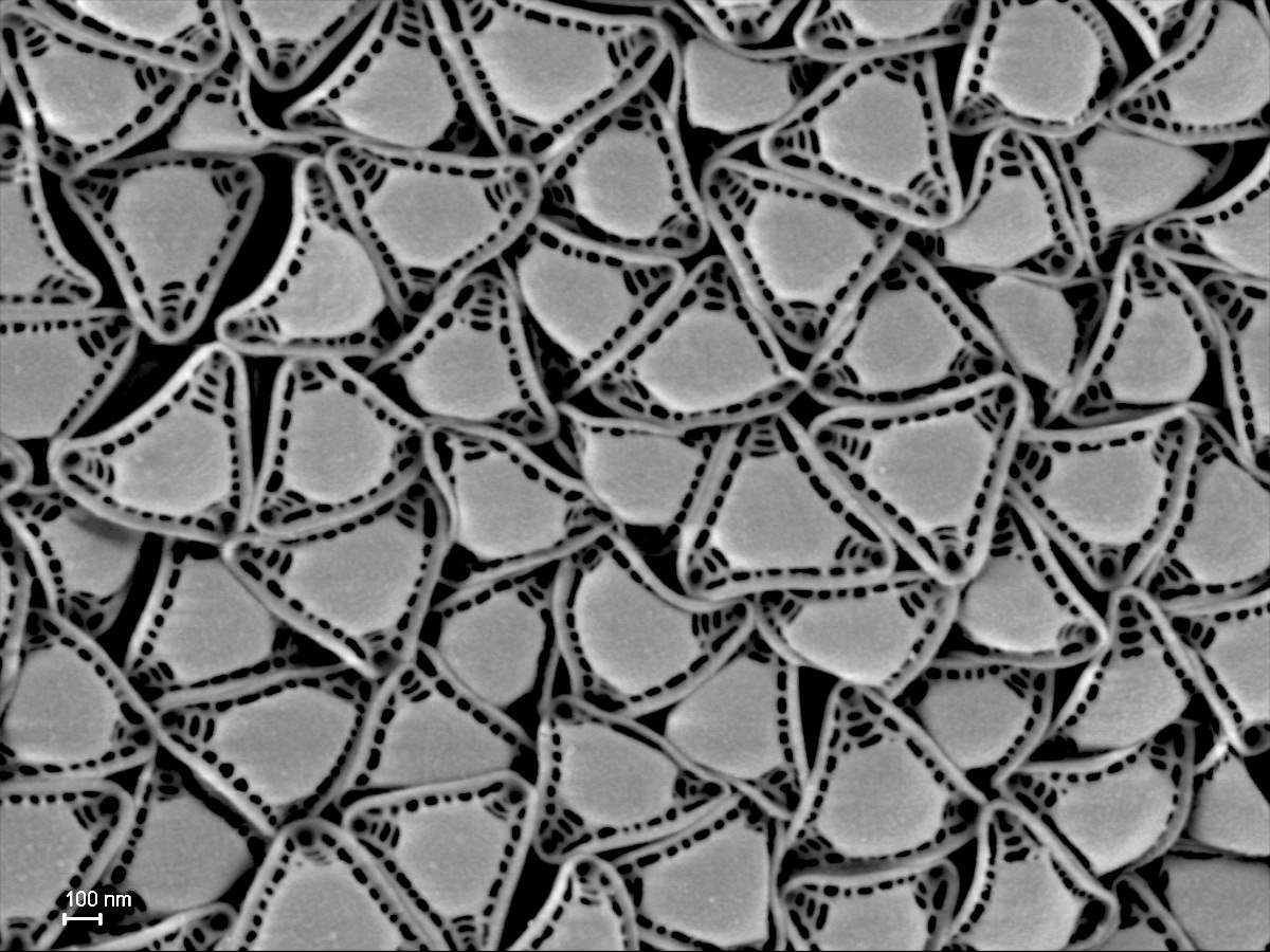
3.1.1.1.1. Pompholyxophrys Archer 1869
Spherical; outer mucilaginous envelope with minute colorless perforated siliceous pearls arranged in concentric layers.
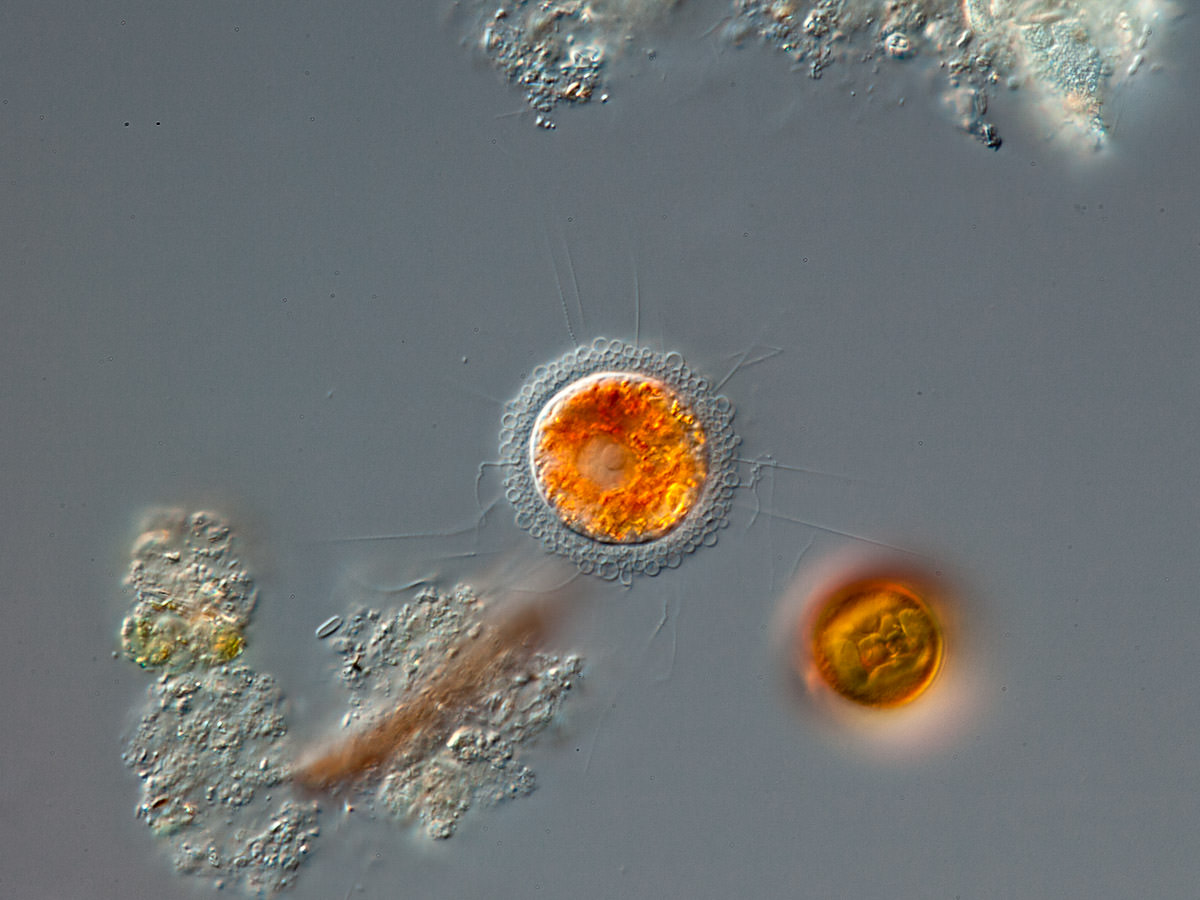
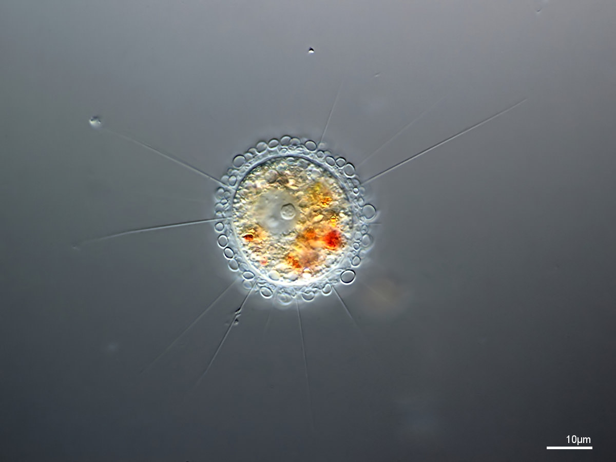
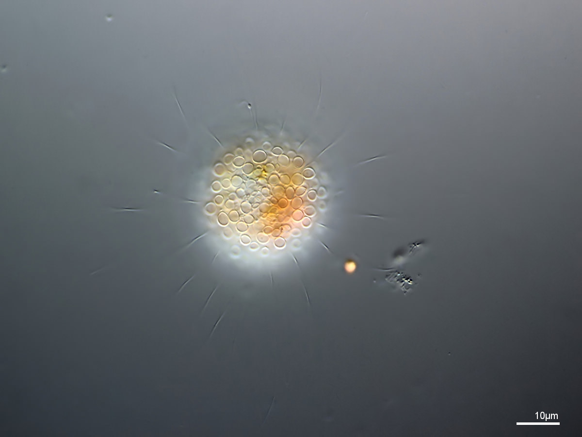
Pompholyxophrys punicea Archer 1869
Pompholyxophrys stellata Nicholls and Dürrschmidt 1985
3.2. Placofila Cavalier-Smith 2012
Heterotroph cells with typically two-tier plate scales, with upper tier usually perforated; non-ciliated with slender radiating usually unbranched filopodia or biciliates with ventral groove emitting branched filopodia.
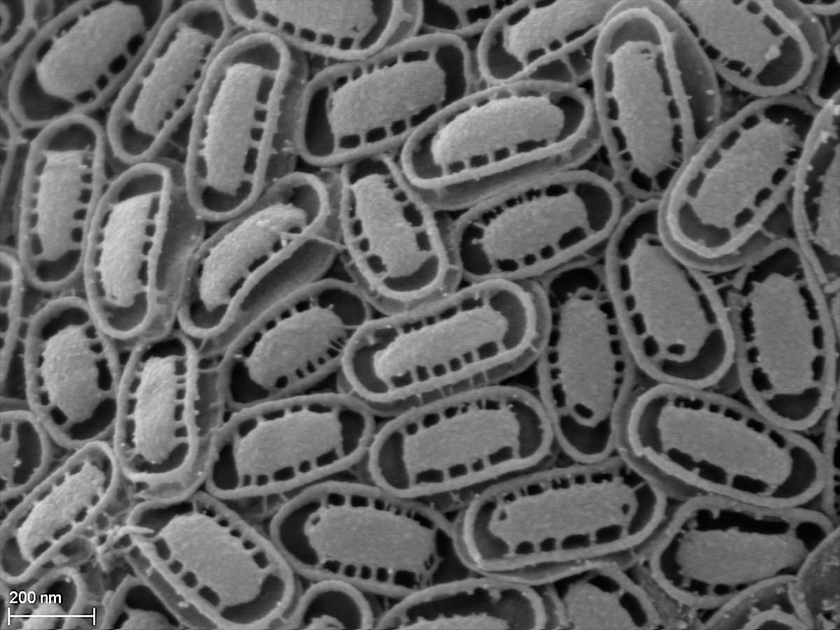
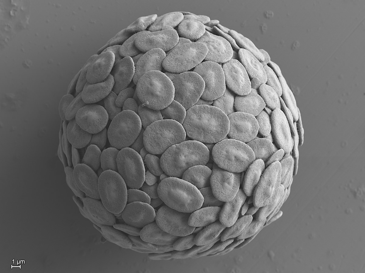
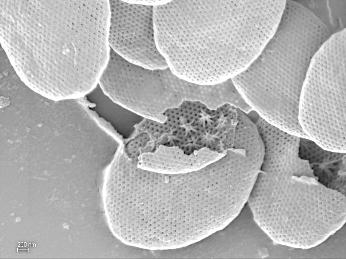
3.2.1. Pinaciophoridae Cavalier-Smith 2012
Non-ciliated heterotrophic uninucleate cells with extremely thin filopodia; tangential two-layered plate scales with numerous perforations in the inner layer.
3.2.1.1. Pinaciophora Greeff 1869
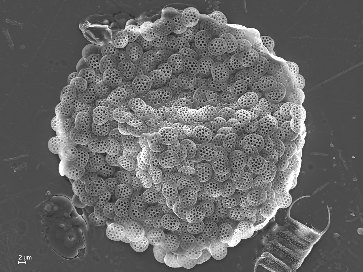
Pinaciophora fluviatilis Greeff 1869